Home

Vânzare Scuipat mal onychomycosis the microscopic image Deplin Fanatic strănut

Figure 1 | Mixed Infection of Toe Nail Caused by Trichosporon asahii and  Rhodotorula mucilaginosa | SpringerLink
Figure 1 | Mixed Infection of Toe Nail Caused by Trichosporon asahii and Rhodotorula mucilaginosa | SpringerLink

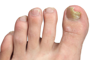
Fungus nail/Onychomycosis - KARR FOOT & LEG CENTER

Nail Fungal Infection Norfolk NE, Norfolk Podiatry Center & Lower Extremity  Nerve Surgery Center
Nail Fungal Infection Norfolk NE, Norfolk Podiatry Center & Lower Extremity Nerve Surgery Center

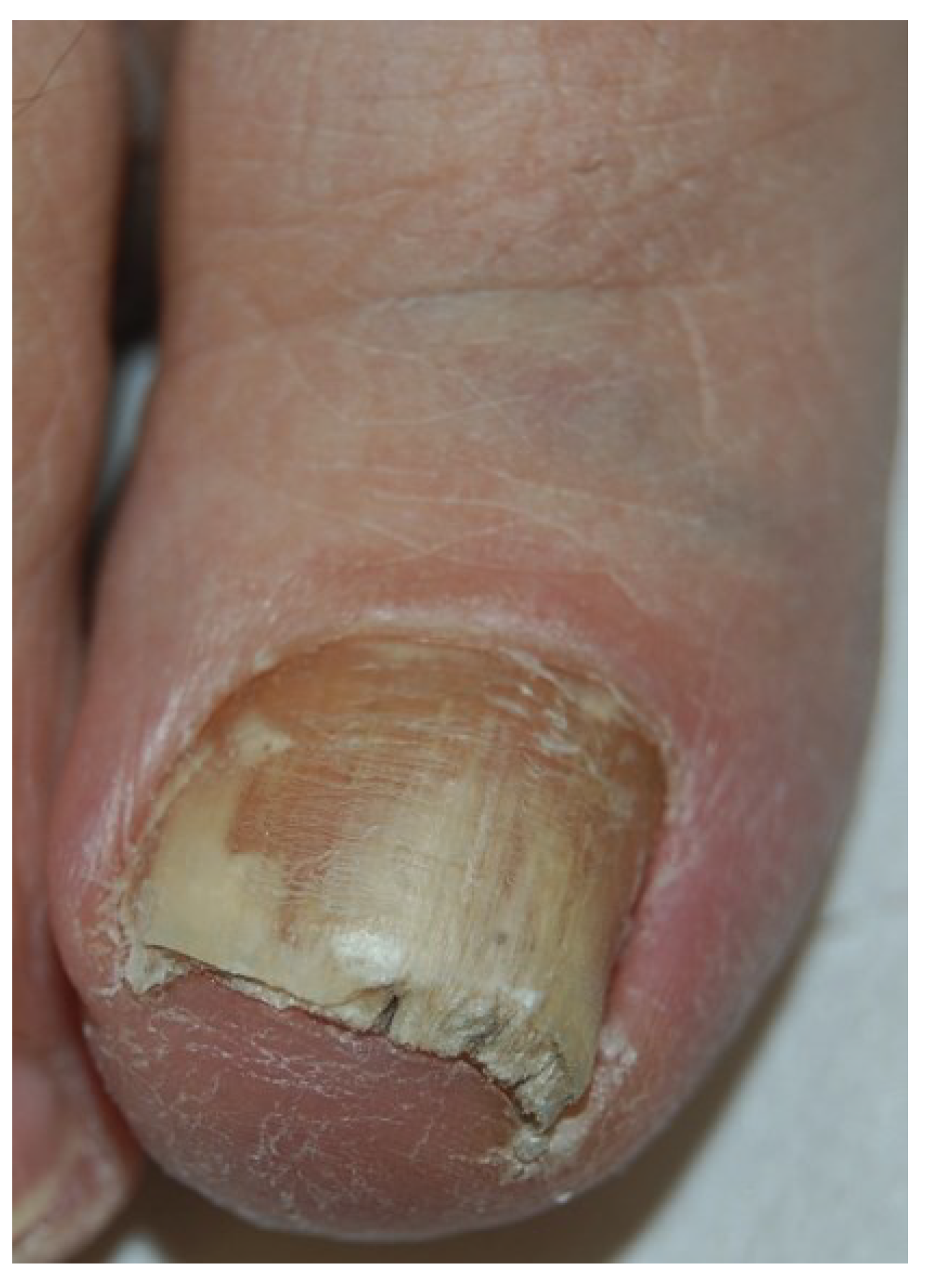
Onychomycosis: Current Trends in Diagnosis and Treatment | AAFP

Fungal Nail Infections (Onychomycosis): A Never-Ending Story? | PLOS  Pathogens
Fungal Nail Infections (Onychomycosis): A Never-Ending Story? | PLOS Pathogens

Frontiers | Diagnosis of Onychomycosis: From Conventional Techniques and  Dermoscopy to Artificial Intelligence
Frontiers | Diagnosis of Onychomycosis: From Conventional Techniques and Dermoscopy to Artificial Intelligence
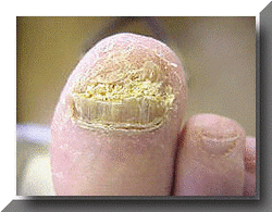
Toe Nail Onychomycosis | cmpt

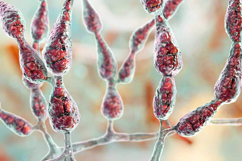
Trichophyton rubrum colony and microscopic appearance (40x). | Download  Scientific Diagram
Trichophyton rubrum colony and microscopic appearance (40x). | Download Scientific Diagram

In Vivo Confocal Microscopy in the Diagnosis of Onychomycosis | MDedge  Dermatology
In Vivo Confocal Microscopy in the Diagnosis of Onychomycosis | MDedge Dermatology

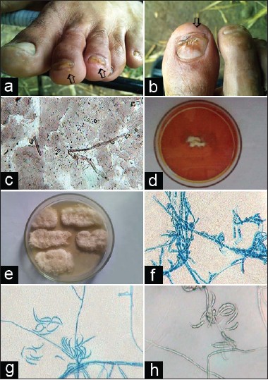
Fusarial onychomycosis among gardeners: A report of two cases - Indian  Journal of Dermatology, Venereology and Leprology
Fusarial onychomycosis among gardeners: A report of two cases - Indian Journal of Dermatology, Venereology and Leprology

JoF | Free Full-Text | Recent Findings in Onychomycosis and Their  Application for Appropriate Treatment
JoF | Free Full-Text | Recent Findings in Onychomycosis and Their Application for Appropriate Treatment

Scanning Electron Microscope Imaging of Onychomycosis

3,388 Onychomycosis Images, Stock Photos & Vectors | Shutterstock

How does a nail fungus look like under Microscope - YouTube

Onychomycosis: Pathogenesis, Diagnosis, and Management | Clinical  Microbiology Reviews
Onychomycosis: Pathogenesis, Diagnosis, and Management | Clinical Microbiology Reviews

Fungal Infected Nail Microscopy showing Hyphae and Chlamydospores of  Dermatophytes - YouTube
Fungal Infected Nail Microscopy showing Hyphae and Chlamydospores of Dermatophytes - YouTube

Scanning Electron Microscope Imaging of Onychomycosis

JoF | Free Full-Text | Onychomycosis: A Review

SciELO - Brasil - Scanning electron microscopy of superficial white  onychomycosis Scanning electron microscopy of superficial white  onychomycosis
SciELO - Brasil - Scanning electron microscopy of superficial white onychomycosis Scanning electron microscopy of superficial white onychomycosis

Onychomycosis: Clinical overview and diagnosis - ScienceDirect

PDF] Scanning electron microscope imaging of onychomycosis. | Semantic  Scholar
PDF] Scanning electron microscope imaging of onychomycosis. | Semantic Scholar

PDF] The Role of Scanning Electron Microscopy in the Direct Diagnosis of  Onychomycosis | Semantic Scholar
PDF] The Role of Scanning Electron Microscopy in the Direct Diagnosis of Onychomycosis | Semantic Scholar

What is Toenail Fungus? Causes & Symptoms of Toenail Fungus

Scanning Electron Microscope Imaging of Onychomycosis